
3D-printed materials are increasingly being used for definitive restorations. But how do their surfaces behave in the oral cavity? Besides strength, aesthetics, and precision, the accumulation of biofilm also determines how well a restorative material performs in the long term. A study has examined this in more detail.
Surface conditioning for the adhesive bonding of dental restorations includes mechanical and chemical pretreatment steps. This study examines both methods and demonstrates their importance for secure and durable adhesive bonding.
The increasing use of additive manufacturing technologies in dentistry opens up new possibilities for the production of individual dental restorations. The layer-by-layer construction in 3D printing enables complex geometries and makes processes such as stereolithography (SLA) and digital light processing (DLP) an attractive option for the production of polymer-based temporary and increasingly also definitive fixed dental prostheses (FDPs) [1].
For the long-term clinical success of 3D-printed restorations, biological aspects are of particular importance in addition to mechanical properties and aesthetic requirements [2]. The surface of a dental restoration represents the direct contact area with the oral environment and significantly influences the adhesion of microorganisms and the development of bacterial biofilms. Extensive biofilm formation can lead to discoloration, gingival irritation, gingivitis, and secondary caries, thus impairing the lifespan and aesthetics of a restoration [3, 4].
The microbial colonization of dental materials is influenced by their chemical composition and surface properties such as roughness and wettability [5]. However, current studies indicate that not roughness alone, but the entire surface structure determines initial biofilm adhesion [6]. While the mechanical properties of 3D-printed dental materials have already been extensively investigated, their behavior towards complex oral biofilms has so far only been insufficiently characterized.
Material and method
To investigate biofilm formation, cylindrical test specimens (Ø 10 mm, thickness 2 mm) were manufactured from the following dental restorative materials and post-processed according to the manufacturer's instructions.
- Additively manufactured 3D printing resins: VSC (VarseoSmile Crown plus), ND (NextDent C&B MFH), VST (VarseoSmile Temp), TP (Temp PRINT), P (P Pro Crown & Bridge)
- Subtractively milled polymer blocks: TC (Tetric CAD = composite), TEL (Telio CAD = PMMA)
- Conventionally processed: TEC (Tetric EvoCeram = photopolymerizing), PT (Protemp 4 = autopolymerizing)
- Control group: Zr (Tizian Blank Translucent Zirconium Dioxide = Zirconium oxide)
After disinfection, the test specimens were incubated with a multispecies biofilm consisting of Actinomyces naeslundii, Streptococcus gordonii, S. mutans, S. oralis, and S. sanguinis for 72 h. The biofilm mass was quantified by crystal violet staining, while the number of viable bacteria was determined by colony counting after biofilm removal. Additionally, surface properties such as roughness, wettability, and interfacial tension were analyzed to assess their influence on bacterial adhesion.
Results
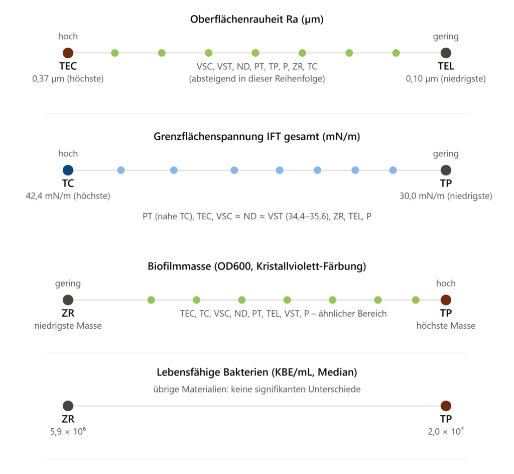
Surface roughness (Ra) varied between the materials, ranging from 0,10 µm (TEL, lowest) to 0,37 µm (TEC, highest), with significant differences primarily observed between subtractively manufactured and 3D-printed specimens. Interfacial tension (IFT) ranged from 30,0 mN/m (TP, lowest) to 42,4 mN/m (TC, highest). TC exhibited significantly higher values than all materials except PT, while TP showed significantly lower values than all materials except PT. The three 3D printing resins, VSC, ND, and VST, were in a similar mid-range, with values of 34,4–35,6 mN/m (Figure 1). Regarding biofilm mass, ZR showed the lowest, and TP the highest. The remaining materials—TEC, TC, VSC, ND, PT, TEL, VST, and P—were in a similar range. The median number of viable bacteria (CFU/mL) ranged from 5,9 × 10⁶ CFU/mL (ZR) to 2,0 × 10⁷ CFU/mL (TP), with no significant differences between the other materials (Figure 1).
Conclusion
The study [7] shows that the susceptibility of 3D-printed materials to biofilm formation does not differ significantly from that of conventionally processed polymer materials. This applies to both the amount of biofilm accumulated and the number of living bacteria within it. Overall, the results suggest that 3D-printed materials represent an alternative for both temporary and permanent dental prostheses.
For practice and laboratory, this means
3D printing does not automatically mean more biofilm. In the in-vitro model studied, the 3D-printed materials did not show significantly greater biofilm formation overall than the conventionally processed polymer materials. At the same time, the individual materials differed in their surface properties and, in some cases, in the amount of biofilm. Therefore, a simple classification based solely on the manufacturing process is insufficient.
examination
Wuersching SN, Manghofer D, Stawarczyk B, Gueth JF, Kollmuss M. Surface Properties of Dental Materials Influence the In Vitro Multi-Species Biofilm Formation. Polymers (Basel) 2026;18(2):288.
Books
[1] Tian Y, Chen C, Xu X, Wang J, Hou X, Li K, Lu X, Shi H, Lee ES, Jiang HB. A Review of 3D Printing in Dentistry: Technologies, Affecting Factors, and Applications. Scanning 2021;9950131.
[2]Wuersching SN, Hickel R, Edelhoff D, Kollmuss M. Initial Biocompatibility of Novel Resins for 3D Printed Fixed Dental Prostheses. Dent Mater 2022;38:1587–1597.
[3] Kolenbrander PE, London J. Adhere Today, Here Tomorrow: Oral Bacterial Adherence. J Bacteriol 1993;175:3247–3252.
[4] Jokstad A. Secondary caries and microleakage. Dent Mater 2016;32:11–25.
[5] Schmalz G, Cieplik F. Biofilms on Restorative Materials. Monogr. Oral Sci 2021;29:155–194.
[6] Hsu LC, Fang J, Borca-Tasciuc DA, Worobo RW, Moraru CI. Effect of Micro- and Nanoscale Topography on the Adhesion of Bacterial Cells to Solid Surfaces. Appl Environ Microbiol 2013;79:2703–2712.
[7] Wuersching SN, Manghofer D, Stawarczyk B, Gueth JF, Kollmuss M. Surface Properties of Dental Materials Influence the In Vitro Multi-Species Biofilm Formation. Polymers (Basel) 2026;18(2):288. https://doi.org/10.3390/polym18020288